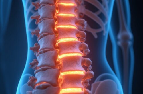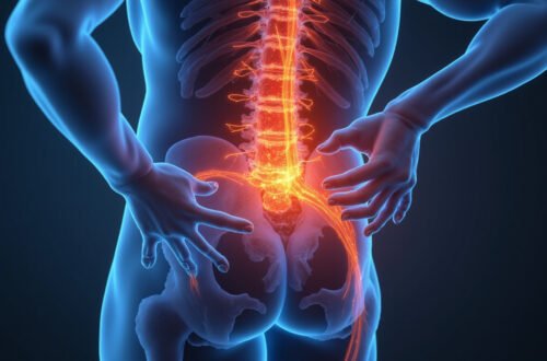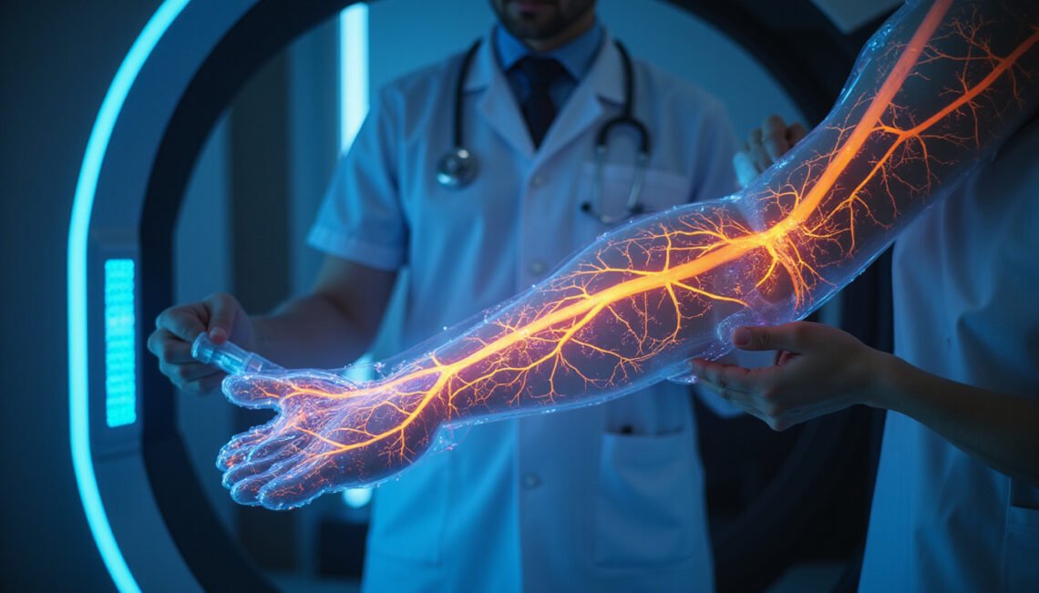
Nerve damage used to be something doctors often inferred indirectly—by symptoms, reflex tests, or slow electrical studies—rather than seeing it. Today, advances in nerve imaging are changing that. From high‑resolution MRI to ultrasound that can see individual nerve fascicles, clinicians can now visualize neuropathy earlier, more precisely, and often less invasively than before.
This article walks through what nerve imaging is, the latest breakthroughs, how it helps detect hidden neuropathy faster, and what patients can expect.
What Is Nerve Imaging?
Nerve imaging refers to any medical imaging technique used specifically to visualize peripheral nerves or nerve pathways in the body. Instead of only looking at bones or muscles, these tools highlight:
- The structure and course of nerves
- Changes in nerve size or shape
- Surrounding tissues that may be compressing or damaging nerves
- Sometimes, indirect signs of nerve inflammation or degeneration
Historically, diagnosis of neuropathy relied mostly on:
- Neurological exam
- Electromyography (EMG)
- Nerve conduction studies (NCS)
- Laboratory tests for causes like diabetes or autoimmune disease
These are still crucial, but they have limits. EMG and NCS can be uncomfortable, and they measure function, not structure. They can also miss early or very focal nerve damage. Nerve imaging fills this gap by helping clinicians literally see what’s happening to the nerves.
Why Faster Detection of Neuropathy Matters
Neuropathy is damage or dysfunction of one or more nerves. It can cause:
- Numbness, tingling, or “pins and needles”
- Burning or shooting pain
- Weakness and muscle wasting
- Balance problems
- Autonomic symptoms (e.g., heart rate, digestion, sweating issues)
Common causes include diabetes, autoimmune disorders, compression (like carpal tunnel), trauma, vitamin deficiencies, infections, and chemotherapy drugs.
Earlier, accurate detection matters because:
- Treatment can begin sooner, potentially preventing permanent damage.
- Reversible causes (like vitamin deficiencies, entrapment syndromes, or inflammatory neuropathies) can be corrected.
- Surgical decisions can be better informed, targeting the exact site of compression or injury.
- Monitoring disease progression becomes more precise, guiding medication adjustments and rehabilitation.
Nerve imaging doesn’t replace nerve conduction testing, but when combined, it offers a much clearer picture—both literally and diagnostically.
Main Types of Nerve Imaging Used Today
Different tools reveal different aspects of nerve health. The main nerve imaging modalities in clinical practice are:
1. High-Resolution Ultrasound of Nerves
Ultrasound is one of the most exciting developments in nerve imaging. Using high-frequency sound waves, clinicians can see nerves in real time.
Key advantages:
- No radiation
- Fast and relatively low cost
- Dynamic: you can watch the nerve move with joints or pressure
- Can be done at the bedside or in a clinic
What doctors can see:
- Nerve cross-sectional area (swelling or thinning)
- Changes in nerve structure (fascicle patterns)
- Entrapment by surrounding tissues (e.g., in carpal tunnel syndrome)
- Cysts, tumors, or scars compressing nerves
- Post-surgical nerve changes
Ultrasound has become particularly valuable for diagnosing entrapment neuropathies (like ulnar nerve compression at the elbow) and for guiding targeted injections around nerves.
2. MRI Neurography (MRN)
Magnetic Resonance Neurography is a specialized form of MRI tailored for nerves. It uses specific sequences to highlight nerve bundles and suppress signals from nearby tissues such as fat and muscle.
Benefits of MR neurography:
- Excellent soft tissue contrast
- Visualizes deep nerves not accessible by ultrasound (e.g., in the pelvis or spine)
- Can show inflammation, edema, and structural damage
- Maps long segments of nerve pathways
MRN is especially helpful for:
- Sciatic nerve problems
- Brachial plexus injuries
- Complex pelvic or spinal nerve pain
- Unexplained limb pain where site of nerve damage is unclear
Research continues to refine MRN techniques, with higher resolution and faster scans making it more practical in everyday clinical use.
3. Diffusion Tensor Imaging (DTI) and Tractography
DTI is an advanced MRI technique that measures the diffusion of water molecules in tissues. Nerves and white matter tracts in the brain and spinal cord have directional water flow along fibers, and DTI can map this.
Use in nerve imaging:
- Visualizes white matter tracts in the brain and spinal cord
- Assesses integrity of nerve fiber bundles (e.g., after trauma or stroke)
- Creates colorful “tractography” maps that show nerve pathways
In peripheral neuropathy, DTI is still more research-oriented but is increasingly used in specialized centers to study microstructural nerve changes before they show up on conventional imaging.
4. CT and CT Myelography
CT scanning uses X-rays and is not typically the first choice for peripheral nerve imaging due to limited soft tissue contrast. However, it’s valuable for:
- Showing bony structures that may compress nerves (e.g., narrowing of spinal canals or foramina)
- CT myelography, where contrast dye is injected into cerebrospinal fluid to outline spinal nerves, especially when MRI is not possible
CT remains important in pre-surgical planning—especially around the spine and joints where both bones and nerves must be considered.
Breakthroughs Revolutionizing Nerve Imaging
Recent developments are turning nerve imaging from a niche into a core diagnostic tool in neurology and pain medicine.
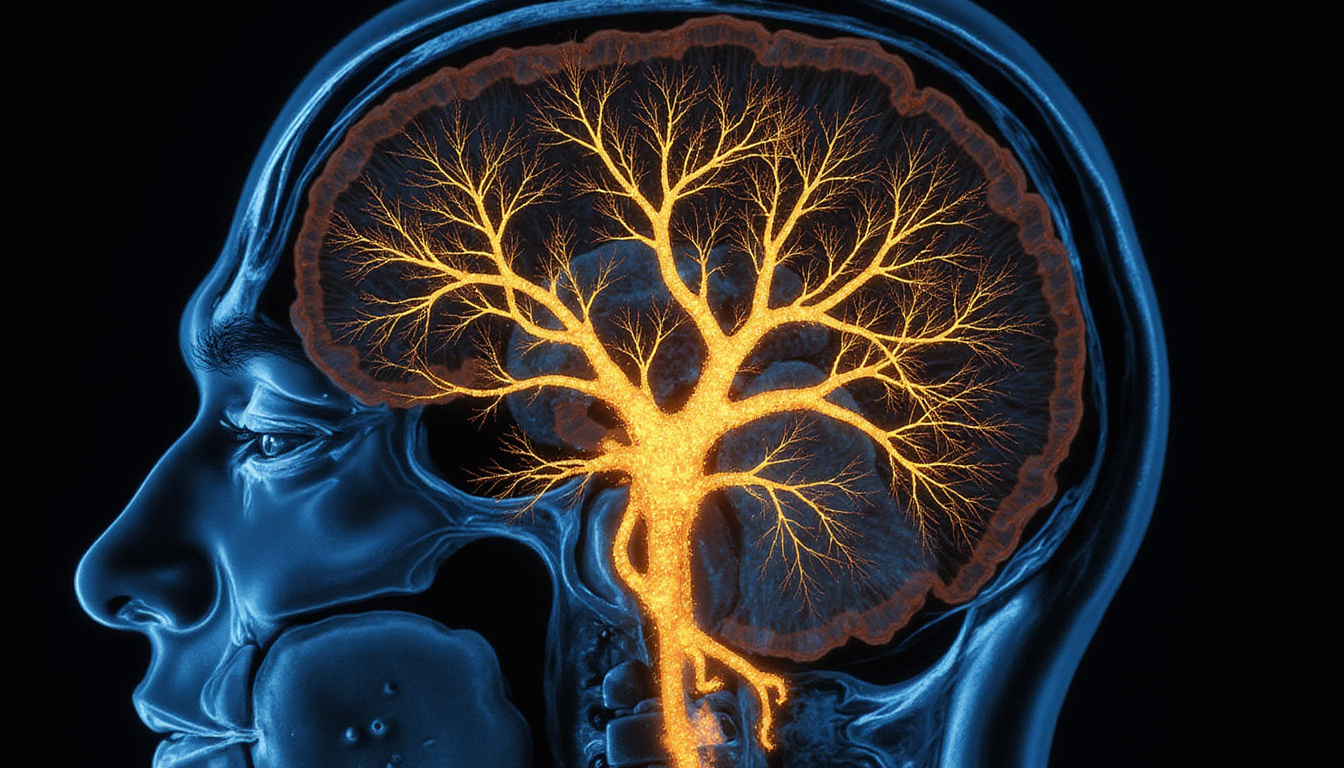
Higher Resolution and Faster Scans
Advances in hardware and software mean:
- Higher-frequency ultrasound probes reveal finer nerve structures.
- 3T and even 7T MRI scanners offer sharper images.
- Faster sequences reduce scan times and motion artifacts.
Result: Doctors see more detail in less time, letting them detect subtle neuropathies earlier.
Quantitative Imaging and AI Assistance
Newer techniques aim to move from “looking” to “measuring” nerves:
- Quantitative MRI can provide metrics like fractional anisotropy or T2 relaxation times, reflecting nerve health.
- Automated segmentation and AI algorithms help highlight abnormal nerve regions, compare to reference values, and track changes over time.
Though much of this is still in advanced centers or research settings, it points to a future where nerve imaging reports contain standardized scores, not just descriptive impressions.
Imaging-Guided Nerve Interventions
Imaging isn’t just diagnostic—it’s becoming therapeutic:
- Ultrasound-guided nerve blocks for pain relief
- Precise steroid or anesthetic injections around compressed nerves
- Image-guided ablation procedures for chronic pain
- Guidance for nerve repair or grafting surgeries
This combination of seeing and treating in real time can shorten the path from diagnosis to relief.
How Nerve Imaging Helps Reveal “Hidden” Neuropathy
“Hidden neuropathy” refers to nerve damage that doesn’t show up clearly on routine tests, or that presents with vague or widespread symptoms. Nerve imaging can uncover:
-
Early or mild entrapments
Ultrasound or MRN can show slight nerve swelling or compression before EMG abnormalities appear. -
Focal lesions in complex areas
MR neurography can identify a small nerve being pinched in the pelvis, shoulder, or deep muscles that would otherwise be missed. -
Non-entrapment causes
Inflammatory changes, tumors, or cysts touching nerves can be directly visualized. -
Post-surgical or post-trauma changes
Imaging can distinguish between a scar binding a nerve, a neuroma (nerve growth at an injury site), or recurrent compression—key in deciding next steps. -
Differences between nerve and muscle problems
By looking at both nerves and surrounding muscles, imaging helps differentiate neuropathy from primary muscle disease.
This detailed visualization can prevent misdiagnoses, reduce unnecessary procedures, and personalize treatment.
What to Expect During a Nerve Imaging Test
The exact experience depends on the modality, but in general:
Ultrasound Nerve Imaging
- Done in a clinic or imaging lab
- You’ll sit or lie down while gel is applied to the skin
- A small transducer is moved over the area of interest
- Usually painless and takes 15–45 minutes
- You may be asked to move joints or change positions during scanning
MRI / MR Neurography
- Performed in a hospital or imaging center
- You lie on a table that slides into the MRI scanner
- Must stay still while loud tapping or thumping sounds occur
- Ear protection and sometimes music are provided
- Typically 30–60 minutes depending on the study
- Not painful, but can be uncomfortable if you dislike enclosed spaces; sedation can be arranged in some cases
CT / CT Myelography
- CT alone is quick (often under 10–15 minutes)
- Myelography involves a lumbar puncture to inject contrast into the spinal fluid, then CT imaging
- You’ll be monitored afterward to reduce post-procedure headache risk
Always tell your provider about implants, pacemakers, pregnancy, or contrast dye allergies before imaging.
When Doctors Recommend Nerve Imaging
Your clinician might suggest a nerve imaging study if:
- Symptoms suggest neuropathy but EMG/NCS are normal or inconclusive
- There’s localized pain, weakness, or numbness along a nerve path
- You have signs of nerve compression (e.g., carpal tunnel, ulnar neuropathy) and treatment decisions depend on severity and exact site
- There’s trauma, surgery, or suspected tumor involving nerves
- Pain persists despite treatment and the cause remains unclear
Nerve imaging is often one piece of a diagnostic puzzle—used in combination with history, physical exam, lab work, and electrodiagnostics.
Benefits and Limitations of Nerve Imaging
Key Benefits
- Earlier diagnosis of some neuropathies
- More precise localization of nerve lesions
- Better surgical and interventional planning
- Monitoring response to therapy over time
- Often noninvasive and relatively safe
Important Limitations
- Not all neuropathies are visible, especially small fiber neuropathies that affect tiny pain fibers in the skin.
- Image quality varies between machines and operators, particularly for ultrasound.
- Some findings may be subtle or nonspecific, requiring expert interpretation.
- Cost and availability can limit access; advanced MR neurography and DTI may be available only at specialized centers.
Professional societies like the American Academy of Neurology provide guidance on when and how to integrate imaging into neuropathy workups (source: AAN Practice Guidelines).
How to Discuss Nerve Imaging With Your Doctor
If you suspect neuropathy or already have a diagnosis, you can ask your provider:
- “Would nerve imaging, such as ultrasound or MR neurography, help clarify what’s causing my symptoms?”
- “Is there a way to see whether a specific nerve is being compressed or inflamed?”
- “If imaging shows the exact site of nerve damage, how would that change my treatment?”
Be prepared to discuss:
- Your complete symptom history
- Any prior imaging, EMG, or lab results
- Medications and medical conditions that affect nerves (e.g., diabetes, autoimmune disease, chemotherapy)
Your clinician will weigh the potential benefits of nerve imaging against factors like cost, availability, and how likely the results are to change management.
FAQ: Nerve Imaging and Neuropathy
Q1: Can nerve imaging detect neuropathy better than nerve conduction studies?
Nerve imaging and nerve conduction studies provide different information. NCS/EMG measure how well nerves function, while nerve imaging shows their structure and surroundings. Often, the best diagnosis comes from combining both—especially for entrapment neuropathies and unexplained focal symptoms.
Q2: Is ultrasound nerve imaging accurate for carpal tunnel and similar conditions?
High-resolution ultrasound nerve imaging is very useful for carpal tunnel syndrome, ulnar neuropathy at the elbow, and other entrapment syndromes. It can show nerve swelling, compression, and adjacent structures. Accuracy depends on the equipment and the operator’s expertise, but in many centers it now complements or sometimes reduces the need for more invasive tests.
Q3: Can MRI nerve imaging show small fiber neuropathy?
Most MRI-based nerve imaging, including MR neurography, is not sensitive enough for pure small fiber neuropathy, which affects tiny pain and temperature fibers. Diagnosis of small fiber neuropathy usually relies on clinical exam, specialized autonomic tests, and skin biopsy rather than imaging. However, MRI can rule out other structural causes that might mimic small fiber symptoms.
Take the Next Step Toward Clarity and Relief
If you’re living with unexplained numbness, tingling, burning pain, or weakness, you don’t have to settle for uncertainty. Modern nerve imaging opens a window into what’s really happening to your nerves—often revealing problems earlier and more clearly than was possible just a decade ago.
Ask your neurologist, pain specialist, or primary care doctor whether nerve ultrasound, MR neurography, or other advanced imaging could sharpen your diagnosis and unlock more targeted treatment options. The sooner the cause of neuropathy is identified, the better your chances of preserving nerve function, relieving pain, and getting back to the life you want to lead.