Electromyography is a diagnostic test that helps doctors understand whether your muscles and the nerves that control them are working properly. If you’ve been experiencing unexplained weakness, numbness, tingling, or muscle cramps, an electromyography (EMG) study may be the key to identifying the source of your symptoms — whether it’s a pinched nerve, a peripheral neuropathy, or a muscle disease.
What electromyography measures and why it matters
Electromyography records the electrical activity produced by skeletal muscles and the nerves that activate them. When a nerve is healthy, it conducts electrical signals smoothly; when a nerve is damaged, those signals change in predictable ways. An EMG lets your clinician see those changes in real time, helping to distinguish between nerve problems (neuropathy), nerve-root problems (radiculopathy), and primary muscle disorders (myopathy).
How an EMG differs from a nerve conduction study
Electromyography typically goes hand-in-hand with nerve conduction studies (NCS). While EMG involves inserting a fine needle electrode into muscles to record electrical activity during rest and contraction, an NCS measures how fast and how strong electrical impulses travel along a nerve using surface electrodes. Together, these tests provide a fuller picture: NCS assesses nerve signal transmission, and EMG evaluates the response of the muscle to that input.
Common reasons doctors order an electromyography
Patients are often referred for EMG when symptoms suggest nerve or muscle involvement. Common indications include:
- Persistent numbness or tingling in the hands or feet
- Unexplained muscle weakness or wasting
- Sudden onset of muscle pain, cramps, or twitching
- Suspected conditions such as carpal tunnel syndrome, peripheral neuropathy, amyotrophic lateral sclerosis (ALS), or myositis
- To localize the level of nerve compression or injury
What to expect during the electromyography test
Knowing what will happen during an EMG can ease anxiety. The test is usually performed in a clinic or hospital electromyography lab and takes 30 to 90 minutes, depending on how many muscles and nerves are tested. Typical steps include:
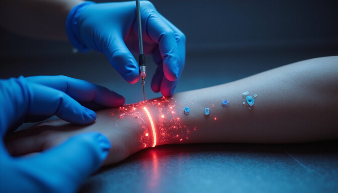
- A clinician places small surface electrodes on the skin for nerve conduction studies.
- For EMG, a thin needle electrode is inserted into several muscles to record electrical activity at rest and during voluntary contraction.
- You may be asked to relax and then to contract the muscle (for example, squeeze a hand or lift a foot) while the clinician records signals.
- The examiner may repeat tests and compare muscles on both sides of the body.
Is electromyography painful?
Discomfort is the most common concern. Needle insertion causes a brief, sharp sensation similar to a vaccine or blood draw, followed by mild aching during muscle contraction. Most people tolerate the procedure without medication. If pain is a major concern, discuss options with your clinician beforehand.
Conditions an EMG can help diagnose
Electromyography is useful for differentiating among several disorders. It can indicate whether symptoms originate in the muscle, the peripheral nerve, the nerve root near the spine, or the neuromuscular junction (where nerve meets muscle). Conditions EMG commonly evaluates include:
- Carpal tunnel syndrome and other entrapment neuropathies
- Peripheral neuropathy from diabetes or toxins
- Radiculopathy from spinal disc herniation
- Motor neuron diseases like ALS
- Myopathies (muscle diseases) such as polymyositis
- Disorders of neuromuscular transmission, like myasthenia gravis
Interpreting EMG results: what the tracings mean
EMG produces characteristic patterns:
- Normal muscles show minimal electrical activity at rest and a clean, coordinated pattern during contraction.
- Denervation (nerve injury) produces spontaneous electrical discharges at rest, called fibrillation potentials and positive sharp waves.
- Chronic nerve injury may show large motor unit potentials from reinnervation, reflecting nerve recovery.
- Myopathic conditions typically show small, short-duration motor unit potentials with early recruitment.
Your clinician integrates EMG findings with your history, physical exam, and other tests (like imaging or blood tests) to arrive at a diagnosis.
Preparing for the test and aftercare
To get accurate electromyography results and minimize issues:
- Avoid lotions, oils, or creams on the day of the test.
- Continue medications unless your provider instructs you otherwise (some medications can alter results).
- Wear loose, comfortable clothing that allows access to the areas being tested.
After the test, mild soreness at needle sites is common for a day or two; applying a warm compress and taking over-the-counter pain relievers usually helps.
Risks and limitations of electromyography
EMG is generally safe, but there are a few potential risks: mild bleeding, bruising, or infection at needle sites (rare) and temporary discomfort. People with bleeding disorders or on blood thinners should tell their clinician in advance. EMG findings also require expert interpretation — an abnormal result doesn’t always pinpoint the underlying cause, and a normal EMG does not rule out every nerve or muscle condition. For comprehensive guidance, organizations like the Mayo Clinic provide reliable overviews (source).
When electromyography won’t give full answers
Some conditions cause intermittent symptoms or small-fiber neuropathies that EMG/NCS may not detect because those tests mainly assess large nerve fibers. In such cases, your clinician may recommend complementary tests like skin biopsy, autonomic testing, MRI, or lab studies.
Practical tips to get the most from your EMG appointment
- Keep a clear symptom diary (when symptoms started, what makes them better or worse).
- Bring a list of medications and medical history.
- Ask your clinician which muscles or nerves will be tested and why.
- Discuss whether you should stop blood thinners or other medications beforehand.
- Bring a friend or family member for support if you’re anxious.
Quick checklist: items to bring and do before an EMG
- Photo ID and insurance information
- Comfortable clothes that allow access to arms or legs
- Medication list and relevant medical records
- Questions prepared for your clinician about the procedure and risks
FAQ: Common patient questions about electromyography
Q: What is an electromyography test and why is it used?
A: An electromyography test records the electrical activity of muscles and nerves to help diagnose nerve damage, muscle disorders, and conditions affecting nerve-to-muscle communication. It’s used when symptoms like weakness, numbness, or twitching suggest a neuromuscular problem.
Q: How does electromyography feel and how long does the test take?
A: The procedure involves small needle insertions that may produce brief discomfort and mild aching during muscle use. Most EMG sessions last between 30 and 90 minutes depending on how many muscles are examined.
Q: Can electromyography detect all nerve problems?
A: Electromyography detects many nerve and muscle disorders, especially those affecting large fibers and motor function, but it may miss small-fiber neuropathies or intermittent problems. Your clinician may combine EMG with other tests for a full evaluation.
When to seek follow-up care
If your EMG shows abnormalities, your clinician will discuss next steps — which could include targeted treatments (physical therapy, medications, injections), surgery for nerve entrapment, or referral to a neuromuscular specialist. If symptoms worsen suddenly (rapidly increasing weakness, severe pain, or new loss of bowel/bladder control), seek urgent medical care.
Authoritative resources
For detailed patient-oriented information, the Mayo Clinic provides a helpful overview of EMG and nerve conduction studies (source): https://www.mayoclinic.org/tests-procedures/emg/about/pac-20393948
Conclusion and call to action
Electromyography is a valuable diagnostic tool that gives clinicians direct insight into how your nerves and muscles are functioning. If you’re living with unexplained weakness, numbness, or muscle twitching, an EMG can be the step that clarifies the cause and points to effective treatment. Talk to your primary care doctor or a neurologist about whether an electromyography study is appropriate for your symptoms. If you’d like, request a referral now or schedule a consultation to review your symptoms and get a personalized plan for diagnosis and care.