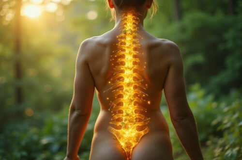
Lumbar MRI: What Your Scan Really Reveals About Back Pain
If you’ve been struggling with back pain, chances are your doctor has mentioned a lumbar MRI or you’re already holding a radiology report that’s full of confusing terms. Understanding what this scan actually shows—and what it doesn’t—can help you make better decisions, reduce anxiety, and choose the right treatment path.
This guide walks you through what a lumbar MRI is, how it works, what your results really mean, and how to use the information wisely alongside your symptoms and exam—not instead of them.
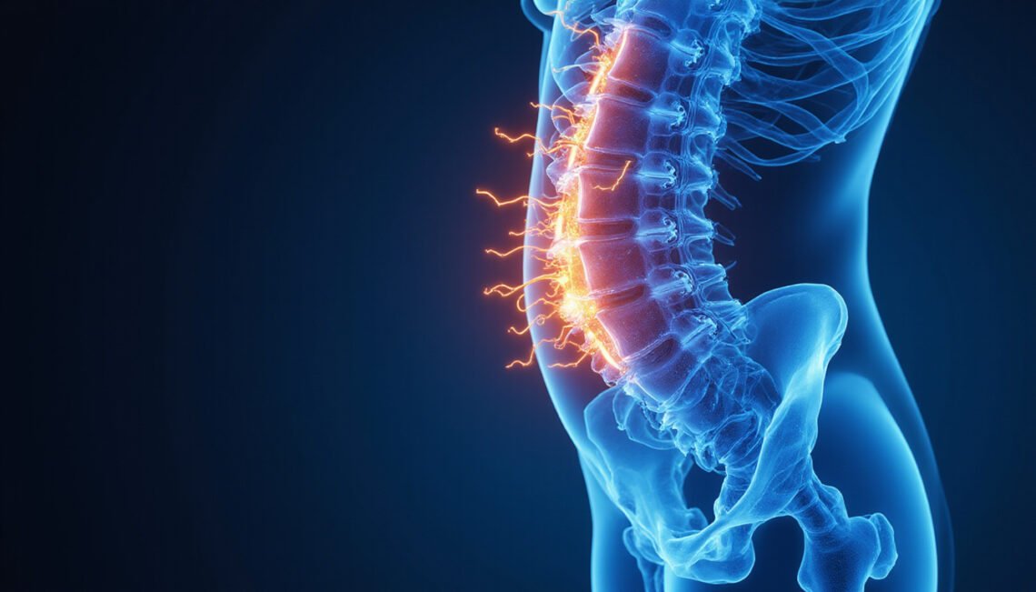
What Is a Lumbar MRI?
A lumbar MRI (magnetic resonance imaging) is an imaging test that uses strong magnets and radio waves to create detailed pictures of your lower spine. Unlike X-rays or CT scans, MRI doesn’t use ionizing radiation.
Your lumbar spine typically includes:
- Five vertebrae (L1–L5)
- Intervertebral discs between each vertebra
- Spinal cord and cauda equina (nerve roots)
- Facet joints
- Ligaments and muscles
- Surrounding soft tissues
A lumbar MRI provides clear images of:
- Discs and their shape/height
- Nerve roots and any compression
- Spinal canal width
- Joints and signs of arthritis
- Soft tissues like ligaments and some muscles
This makes it valuable for evaluating certain causes of back and leg pain, numbness, or weakness.
When Is a Lumbar MRI Actually Needed?
Contrary to popular belief, not everyone with back pain needs a lumbar MRI. In many cases, simple mechanical low back pain improves with time, movement, and basic treatment.
Doctors typically consider ordering a lumbar MRI when:
-
Red flag symptoms are present, such as:
- Severe or rapidly worsening weakness
- Loss of bowel or bladder control
- Numbness in the “saddle” area
- History of cancer, significant trauma, unexplained weight loss, or infection signs (fever, chills)
-
Pain persists beyond 6–8 weeks despite appropriate treatment:
- Physical therapy
- Medication
- Activity modification
-
There’s significant leg pain or neurologic symptoms, such as:
- Sciatica (radiating leg pain)
- Numbness or tingling following a nerve pattern
- Foot drop or other motor weakness
-
Surgery or injections are being considered:
- To guide targeted procedures
- To confirm structural issues likely to respond to intervention
Clinical guidelines from organizations like the American College of Physicians emphasize that imaging for low back pain is usually unnecessary in the first 4–6 weeks unless serious conditions are suspected (source: American College of Physicians).
What Your Lumbar MRI Really Shows
A lumbar MRI can reveal a long list of findings. Some sound scary, but many are simply signs of normal aging, not necessarily the source of your pain.
Common terms you may see:
- Disc bulge – The disc extends slightly beyond its normal boundary.
- Disc herniation – Part of the disc’s inner material pushes out, sometimes compressing a nerve.
- Degenerative disc disease – Age-related disc dehydration and height loss.
- Facet arthropathy – Arthritis in the small joints at the back of the spine.
- Spinal stenosis – Narrowing of the spinal canal or nerve exit holes.
- Spondylolisthesis – One vertebra slips forward over another.
- Annular tear – A small tear in the outer disc ring.
The key is not simply whether these things are present, but:
- Where they are located
- Which nerve roots (if any) they affect
- Whether they match your symptoms and physical exam
Normal Age-Related Changes vs. Problem Findings
One of the most important truths about lumbar MRI is this: many “abnormal” findings are normal for your age and may be painless.
Research has repeatedly shown:
- Many people with no back pain at all have disc bulges, herniations, and degenerative changes on MRI.
- The likelihood of seeing degenerative disc disease and mild stenosis increases with age, even in healthy, active individuals.
In other words, your MRI might look “worse” than how you actually feel.
Normal age-related changes often include:
- Mild disc dehydration (loss of disc height)
- Small disc bulges without clear nerve compression
- Mild facet joint arthritis
- Slightly narrowed disc spaces
These changes alone don’t guarantee pain. They become clinically important when:
- They line up exactly with your pain pattern (e.g., L5 nerve root compression plus L5 symptoms).
- Symptoms are persistent, severe, or progressive.
- They impair function (walking, standing, working, sleeping).
Matching MRI Findings to Your Symptoms
To understand what your lumbar MRI really means, it must be interpreted alongside:
- Your pain location and character
- What aggravates or eases your pain
- Any numbness, tingling, or weakness
- Your physical exam (reflexes, strength, sensation, movement tests)
For example:
-
Disc herniation at L4–L5
- May cause pain down the outer leg, into the foot
- Could produce numbness and weakness with dorsiflexion (lifting the foot)
- If the MRI shows a large herniation compressing the L5 nerve and your symptoms match, the finding is likely significant.
-
Mild bulge at multiple levels
- If there’s no clear nerve compression and your primary problem is localized low back ache with good strength and sensation, these may be incidental findings.
A good spine specialist or radiologist doesn’t just read the pictures—they integrate the images with your clinical story.
Common Misconceptions About Lumbar MRI
Understanding what a lumbar MRI can and cannot do helps you interpret results more calmly and accurately.
-
“If my MRI is normal, my pain isn’t real.”
False. Many causes of back pain—muscle strain, ligament sprain, facet joint irritation, or even some nerve issues—may appear subtle or invisible on MRI. -
“If my MRI is abnormal, I must need surgery.”
Not necessarily. Many disc herniations and degenerative changes improve with non-surgical care. Surgery is usually reserved for:- Severe or progressive neurological deficits
- Confirmed nerve compression with disabling symptoms
- Failure of conservative treatment
-
“The worst-looking MRI equals the worst pain.”
Not true. Some people with alarming-looking scans report little discomfort, while others with modest changes have severe pain. -
“MRI can pinpoint exactly why I hurt.”
MRI is an important piece of the puzzle, but it doesn’t explain everything. Pain can be influenced by:- Muscles and fascia
- Posture and movement habits
- Stress, sleep, and overall health
- Central sensitization (nervous system becoming more sensitive to pain)
How a Lumbar MRI Affects Your Treatment Plan
Once you have lumbar MRI results, your provider will usually combine them with your clinical picture to decide next steps.
Non-Surgical Options (Most Common)
For many people, treatment starts with conservative measures:
- Physical therapy
- Core and hip strengthening
- Flexibility and mobility work
- Posture and movement training
- Medications
- Short-term NSAIDs (if appropriate)
- Occasionally nerve pain medications
- Manual therapy
- Spinal manipulation or mobilization
- Soft tissue treatment
- Lifestyle and ergonomic changes
- Improving workstation setup
- Activity pacing and graded return to exercise
- Pain management techniques
- Heat/ice, TENS, relaxation/breathing strategies
If MRI shows a disc issue or mild stenosis without severe nerve damage, these strategies often provide meaningful relief over time.
Interventional and Surgical Options
If symptoms are severe, persistent, or clearly linked to nerve compression, your team may consider:
- Epidural steroid injections
- To reduce inflammation around irritated nerve roots
- Nerve blocks or facet joint injections
- To confirm and treat certain pain generators
- Surgery
- Microdiscectomy for significant disc herniation with nerve compression
- Laminectomy for severe spinal stenosis
- Fusion for instability or certain advanced conditions
The decision to move toward these options should be based on:
- MRI findings that match your symptoms
- Duration and severity of pain
- Impact on function and quality of life
- Your preferences and health status
Making Sense of Your Lumbar MRI Report
Radiology reports are full of technical language. Here’s how to approach them:
-
Don’t interpret it in isolation.
Always review the report with your doctor, PA, NP, or specialist. -
Ask specific questions:
- Which findings are most likely related to my symptoms?
- Are any of these changes simply age-related?
- Is there any sign of serious or emergency-level problems?
-
Focus on the summary/impression section.
This typically contains the radiologist’s key takeaways. -
Remember that “abnormal” doesn’t always mean “dangerous.”
Words like “degenerative,” “mild,” or “stable” often describe common changes.
Practical Tips Before and After a Lumbar MRI
Here’s a quick checklist to get the most out of your lumbar MRI experience:
-
Clarify the goal.
Ask your provider: “What are we hoping to learn from this lumbar MRI? How will it change my treatment?” -
Bring your symptom history.
Note when pain started, where it goes, what makes it better or worse. -
Ask for plain-language explanations.
After the scan, request a summary in terms you understand. -
Get copies of your images and report.
Helpful if you seek a second opinion or move between providers. -
Avoid catastrophizing.
MRI findings rarely mean your back is “broken” or “ruined.” Many issues are manageable and compatible with an active life.
FAQ About Lumbar Spine MRI and Back Pain
1. Is an MRI of the lumbar spine always necessary for back pain?
No. Most cases of uncomplicated low back pain improve without imaging. A lumbar spine MRI is usually reserved for red flag symptoms, persistent pain beyond several weeks, significant nerve symptoms, or when surgery/injections are being considered.
2. What does a lumbar spine MRI with contrast show that a regular MRI doesn’t?
A lumbar spine MRI with contrast (using gadolinium dye) can highlight inflammation, tumors, infections, or postoperative scar tissue more clearly. It’s not routinely needed for straightforward back pain or typical disc herniations; it’s used when your doctor suspects specific conditions or wants more detailed information.
3. Can a lumbar spine MRI without contrast miss something important?
A lumbar spine MRI without contrast is usually sufficient to detect common problems like disc herniations, stenosis, and many degenerative changes. Contrast is only added if there’s concern for infections, tumors, prior surgery scarring, or certain inflammatory conditions, based on your history and exam.
Use Your Lumbar MRI as a Tool, Not a Verdict
A lumbar MRI can be incredibly useful—but only when it’s ordered for the right reasons and interpreted in the right context. It can clarify the cause of nerve-related leg pain, rule out serious conditions, and guide targeted treatment. At the same time, it can reveal age-related “abnormalities” that may not be causing your pain at all.
If you already have your results, your next best step is to sit down with a knowledgeable clinician who can connect the dots between your imaging, your symptoms, and your goals. If you’re considering whether to get a lumbar MRI, discuss with your provider how the scan’s findings would actually change your care.
Don’t let a report full of medical jargon define your future. Use your lumbar MRI as one piece of a comprehensive plan to understand your back pain, reclaim your mobility, and get back to the activities that matter most to you. If you’re unsure where to start, schedule a visit with a spine-focused physical therapist or physician today and bring your report with you—they can help you turn those images into a clear, actionable path forward.