If you’re dealing with sharp, shooting leg pain, numbness, or tingling, you’ve probably heard the term “sciatica.” What most people don’t realize is how quickly a sciatic nerve ultrasound can help pinpoint the exact source of that pain—and often faster than other tests. Understanding how and when to use this tool can speed up diagnosis, guide better treatment, and help you get back to normal life sooner.
What Is the Sciatic Nerve—and Why Does It Hurt?
The sciatic nerve is the largest nerve in your body. It starts in your lower back, runs through the buttock, down the back of your thigh, and branches all the way to your foot. When something irritates, compresses, or inflames this nerve, you can feel:
- Sharp, burning, or electric pain
- Numbness or tingling
- Weakness in the leg or foot
- Pain that worsens with sitting, bending, or coughing
Common causes include herniated discs, spinal stenosis, piriformis syndrome, injuries, and post-surgical scarring. The challenge is figuring out where along the nerve the problem is. That’s where sciatic nerve ultrasound comes in.
What Is a Sciatic Nerve Ultrasound?
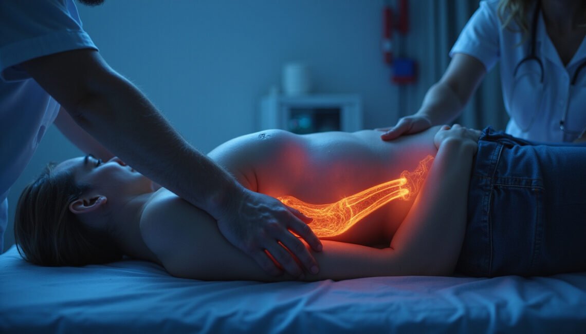
A sciatic nerve ultrasound is a non-invasive imaging test that uses high-frequency sound waves to create real-time pictures of the sciatic nerve and the surrounding tissues. Instead of just looking at bones (like X-rays) or the spine (like MRI), this test directly visualizes the nerve itself as it travels through the buttock and down the leg.
During the exam, a trained clinician:
- Applies gel to your skin.
- Moves a handheld probe (transducer) along the path of the sciatic nerve.
- Views live images of the nerve, muscles, tendons, and nearby structures on a monitor.
No radiation is involved, and the test is usually painless.
Why Use Ultrasound for Sciatica? Key Advantages
The sciatic nerve runs through soft tissues—muscles, fat, fascia—that are not well seen on X-ray. Sciatic nerve ultrasound offers unique advantages in this setting:
1. Direct visualization of the nerve
Ultrasound can show:
- Nerve thickness and swelling
- Changes in nerve structure
- Compression by nearby muscles or masses
- Post-surgical scarring around the nerve
This helps distinguish between nerve entrapment in the buttock or thigh versus a spine-related issue.
2. Dynamic, real-time imaging
Unlike static imaging, ultrasound allows the clinician to:
- Move your leg and hip during the scan
- See how the nerve glides with motion
- Observe nerve impingement that appears only in certain positions
This is especially helpful for conditions like piriformis syndrome, where the nerve’s relationship to the muscle changes with movement.
3. Faster, more accessible than MRI
In many clinics, an ultrasound exam can be performed:
- The same day as your visit
- At a lower cost than MRI
- With no need for a closed, noisy scanner (useful for people with claustrophobia or metal implants that limit MRI)
Ultrasound can rapidly narrow down the problem and determine whether more advanced imaging is truly necessary.
4. Guidance for targeted treatments
One of the biggest advantages is that sciatic nerve ultrasound can guide:
- Precise injections around the nerve
- Hydrodissection (injecting fluid to free a trapped nerve)
- Platelet-rich plasma (PRP) or other regenerative therapies
- Biopsies of suspicious masses adjacent to the nerve
When the clinician sees exactly where the nerve is being compressed, treatment can be delivered to the right spot the first time.
Sciatic Nerve Ultrasound vs. MRI: Which Is Better?
You don’t necessarily have to choose one or the other, because both can be complementary.
MRI excels at:
- Visualizing the spine and discs
- Detecting herniations compressing nerve roots in the back
- Identifying spinal stenosis or tumors within the spinal canal
Ultrasound excels at:
- Imaging the nerve along its course through the buttock and leg
- Evaluating soft tissue causes of sciatica outside the spine (muscles, tendons, cysts, scars)
- Providing dynamic, real-time assessment
Research suggests that musculoskeletal ultrasound is highly accurate for peripheral nerve entrapments and can match or even exceed MRI for some nerve-related conditions when performed by experienced clinicians (source: National Center for Biotechnology Information).
Often, your provider will use sciatic nerve ultrasound to evaluate pain that doesn’t clearly originate from the spine, or when MRI findings don’t fully explain your symptoms.
What Conditions Can Sciatic Nerve Ultrasound Detect?
A well-performed sciatic nerve ultrasound can help uncover several causes of leg pain, including:
- Piriformis syndrome: Compression of the sciatic nerve by the piriformis muscle in the buttock.
- Hamstring injuries: Tears, scarring, or tendinopathy near the ischial tuberosity where the sciatic nerve runs close to the hamstring origin.
- Nerve entrapment by scar tissue: After surgery or trauma.
- Muscle hypertrophy or spasm: That squeezes the nerve.
- Benign or malignant masses: Such as lipomas, cysts, or tumors along the nerve path.
- Post-injection nerve injuries: From injections placed too close to the sciatic nerve in the buttock.
- Neuromas: Thickened, painful nerve segments after injury.
It can also confirm that your sciatic nerve appears normal in size and structure, which may direct attention back to the spine or other causes of leg pain, like vascular disease.
What to Expect During a Sciatic Nerve Ultrasound
Understanding the process can reduce anxiety and help you prepare.
Before the exam
- No special preparation is usually required.
- Wear loose, comfortable clothing; you may need to change into a gown.
- Inform the clinician about your symptoms, prior surgeries, and any implants.
During the exam
The typical steps are:
- Positioning: You may lie on your stomach, side, or back, depending on the area being examined.
- Gel application: A cool gel is applied along the buttock and back of the thigh.
- Scanning: The provider moves the probe along the known path of the sciatic nerve:
- Near the lower back and buttock
- Around the hip
- Down the posterior thigh, possibly to the knee level
- Dynamic testing: You may be asked to bend or rotate your hip or knee to see how the nerve moves.
The exam usually takes 15–30 minutes. You might feel mild pressure from the probe but no significant pain.
After the exam
- You can return to normal activities immediately.
- A radiologist, physiatrist, or specialist trained in musculoskeletal ultrasound will interpret the images.
- Results are typically available quickly and can guide next steps in your care.
How Sciatic Nerve Ultrasound Helps Find the Root Cause Fast
When you’re in pain, time matters. A targeted sciatic nerve ultrasound can accelerate diagnosis in several ways:
-
Quickly rules in or out peripheral nerve problems
If the nerve is clearly swollen or compressed outside the spine, treatment can focus there without delay. -
Clarifies mixed or confusing symptoms
Sometimes MRI shows mild disc bulges that aren’t actually causing your pain. Ultrasound can confirm whether the problem is really in the buttock or thigh instead of the back. -
Reduces trial-and-error treatment
Rather than trying multiple blind injections or medications, ultrasound-guided interventions can be directed at the exact site of compression. -
Identifies red flags
Masses or unusual findings on ultrasound can prompt urgent MRI, CT, or biopsy, helping catch serious problems earlier.
By shortening the path between “something hurts” and “here’s why—and what to do about it,” ultrasound can save weeks or months of uncertainty.
Who Should Consider a Sciatic Nerve Ultrasound?
You might benefit from this test if:
- You have classic sciatica symptoms, but your spinal MRI is normal or inconclusive.
- Your pain worsens with sitting, climbing stairs, or hip movement—suggestive of piriformis or deep gluteal syndromes.
- You’ve had hip or buttock surgery, injections, or trauma, and pain started afterward.
- You have recurrent “hamstring” pain that hasn’t responded to typical treatment.
- You are considering a targeted nerve injection or procedure and want precise guidance.
It’s appropriate to ask your doctor or physical medicine specialist if sciatic nerve ultrasound could provide additional clarity in your particular case.
Limitations and When Ultrasound Isn’t Enough
While powerful, sciatic nerve ultrasound isn’t perfect:
- It cannot see inside the spinal canal as well as MRI.
- Very deep structures or obese body habitus can reduce image quality.
- Accuracy depends heavily on the experience of the person performing the exam.
- Some tiny lesions or early nerve changes may still be missed.
For these reasons, ultrasound is often part of a multimodal approach—used alongside physical exam, MRI, nerve conduction studies, and EMG (electromyography) when necessary.
If you have red-flag symptoms like significant weakness, loss of bladder or bowel control, or rapidly worsening pain, urgent evaluation with MRI and specialist consultation is critical.
How to Prepare and What to Ask Your Provider
To make the most of your sciatic nerve ultrasound appointment:
- Bring prior imaging reports (X-ray, MRI, CT).
- Make a list of:
- When your pain started
- What aggravates or relieves it
- Any previous surgeries or injections
- Wear or bring shorts to allow easy access to the thigh and buttock.
Consider asking:
- “What specific part of the sciatic nerve are you most concerned about?”
- “How will the ultrasound results change my treatment plan?”
- “If the ultrasound is normal, what is the next step in evaluation?”
Clear communication ensures the exam is focused and clinically useful.
FAQ: Sciatic Nerve Ultrasound and Related Tests
1. Is sciatic nerve ultrasound better than MRI for sciatica?
Neither is universally “better.” MRI is superior for detecting disc herniations and spinal stenosis, while sciatic nerve ultrasound is better for visualizing the nerve and soft tissues along the buttock and leg. In many cases, they complement each other to give a complete picture.
2. How much does a sciatic nerve sonography test typically cost?
Costs vary widely by region and facility. In general, sciatic nerve sonography is less expensive than MRI and may be covered by insurance when ordered for appropriate indications. Your imaging center or insurer can provide exact pricing for sciatic nerve ultrasound imaging in your area.
3. Can ultrasound detect all types of sciatic nerve compression?
Ultrasound can detect many forms of peripheral nerve compression—such as piriformis syndrome, muscular entrapment, or masses—but it cannot fully visualize compression occurring inside the spinal canal. If ultrasound of the sciatic nerve is normal yet symptoms persist, further testing like MRI or nerve conduction studies may be needed.
Take Control of Your Sciatica: Don’t Wait on a Diagnosis
Persistent leg pain, numbness, or weakness isn’t something to “wait out,” especially when it interferes with walking, work, or sleep. A sciatic nerve ultrasound is a fast, safe, and often underused tool that can zero in on the true source of your symptoms—whether that’s a tight muscle in the buttock, scarring around the nerve, or a deeper problem that needs urgent attention.
If you’ve been living with sciatica, have unclear MRI findings, or are stuck in a cycle of trial-and-error treatments, talk to your doctor or a musculoskeletal specialist about adding sciatic nerve ultrasound to your evaluation. The right imaging, at the right time, can turn a frustrating mystery into a clear plan—and move you one step closer to lasting relief.