Ischiofemoral impingement is a lesser-known cause of deep buttock and hip pain, often misdiagnosed as sciatica, hamstring strain, or simple “low back pain.” Because it doesn’t show up clearly without targeted imaging and a careful exam, many people go months or years without real answers. Understanding what ischiofemoral impingement is, why it happens, and how to treat it can help you take control of your pain and get back to normal life faster.
What Is Ischiofemoral Impingement?
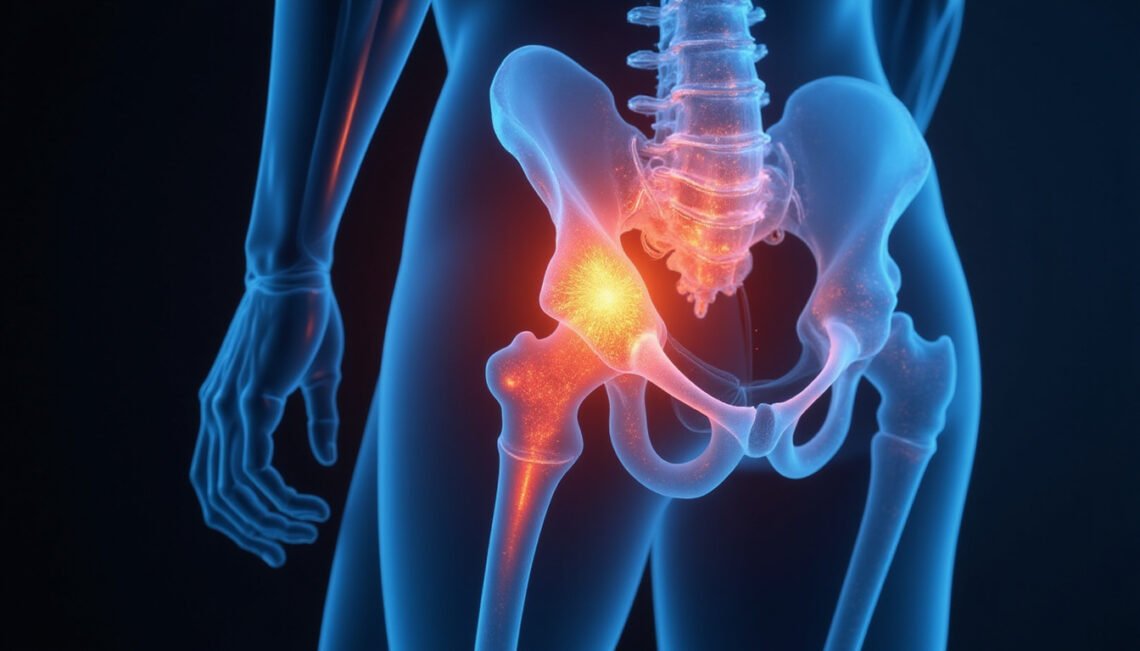
Ischiofemoral impingement (IFI) is a condition where the space between two bony structures in your pelvis and hip—the ischium (part of your pelvis where you “sit”) and the femur (thigh bone)—becomes too narrow.
Specifically, the impingement occurs in the ischiofemoral space, the area between:
- The ischial tuberosity (the “sit bone”)
- The lesser trochanter (a bony bump on the inner part of your upper thigh bone)
Running through this narrow space is the quadratus femoris muscle, a small but important muscle that helps rotate your hip and stabilize your pelvis. When the space is reduced, the quadratus femoris can be compressed, irritated, or damaged, causing pain in the buttock and hip, sometimes radiating down the leg.
Who Is Most at Risk?
Although ischiofemoral impingement can affect anyone, certain groups are more prone:
- Middle-aged women – Several studies suggest a higher prevalence in women, possibly due to pelvic shape and hip alignment differences (source: National Library of Medicine).
- Post-surgical hip patients – Particularly those who have had:
- Total hip replacement
- Hip fracture surgery
- Osteotomy (surgical bone realignment)
- Athletes – Runners, dancers, and athletes who perform repetitive hip extension and rotation.
- People with prior hip trauma or deformity – Fractures, dislocations, or congenital hip issues can alter bone positioning and narrow the ischiofemoral space.
Surprising Causes of Ischiofemoral Impingement
The causes of ischiofemoral impingement can be structural, postural, or activity-related. Some are obvious, but others are surprisingly subtle.
1. Changes After Hip Surgery
Hip surgeries can alter the alignment of the femur and pelvis:
- A slightly lengthened or rotated femur after total hip replacement
- A metal implant that changes the shape or position of the bone
- Scar tissue or changes in soft tissues around the hip
Even small alterations can decrease the ischiofemoral space enough to compress the quadratus femoris muscle.
2. Pelvic Shape and Alignment
Your natural anatomy plays a major role:
- A wider pelvis, more common in women, can change the angle between the femur and the pelvis.
- Femoral version (the twist in your thigh bone) may make certain hip positions more likely to cause impingement.
- Leg length discrepancy can shift pelvic tilt and cause one side to impinge more than the other.
These are usually not “abnormal” on their own, but in combination with repetitive movement or injury, they can cause ischiofemoral impingement symptoms.
3. Repetitive Hip Extension Activities
Movements that repeatedly take the leg backward (hip extension) and rotate it outward can narrow the ischiofemoral space:
- Distance running and sprinting
- Dance (especially ballet, modern, and jazz)
- Gym exercises like lunges, step-ups, and certain glute-focused moves
- Sports that involve long strides or kicking
Over time, this can irritate the quadratus femoris and surrounding tissues.
4. Previous Hamstring or Glute Injuries
Chronic or poorly healed injuries in the back of the thigh or buttock can:
- Alter the way you move
- Change muscle activation patterns
- Increase tension in adjacent muscles and fascia
These compensations may subtly shift the hip and pelvis in ways that promote ischiofemoral impingement.
5. Prolonged Sitting and Poor Posture
Prolonged sitting, especially in positions that tilt the pelvis backward or cross the legs tightly, can:
- Compress the deep gluteal region
- Irritate the quadratus femoris over time
- Reduce the natural mobility of the hip joint
For some people, simply the combination of long hours at a desk and weekend athletics can be enough to trigger IFI.
Key Symptoms: How Ischiofemoral Impingement Feels
The symptoms of ischiofemoral impingement can closely mimic other hip and back problems, which is why it’s often overlooked.
Common symptoms include:
- Deep buttock pain
- Usually on one side
- Felt low in the buttock, near the crease, or slightly to the inner side
- Pain in the back or inner side of the upper thigh
- Sometimes mistaken for hamstring strain
- Pain that worsens with:
- Long strides (walking fast or running)
- Taking the leg backward (hip extension), e.g., going upstairs, pushing off when walking
- External rotation (turning your toes outward)
- Lying on your side with the top leg extended backward
- Tenderness deep in the gluteal area
- Pressing just inside the “sit bone” can reproduce pain
- Occasional radiation down the leg
- Often confused with sciatica, but usually without strong numbness or tingling
Many people say the pain is hard to “pinpoint” and feels deep and aching, sometimes sharp with certain movements.
How Ischiofemoral Impingement Is Diagnosed
Diagnosis relies on a combination of history, physical exam, and imaging.
Clinical Examination
A knowledgeable clinician (sports medicine doctor, orthopedic specialist, or experienced physical therapist) will:
- Ask about activities, previous injuries, and surgeries
- Check hip range of motion
- Try to reproduce your symptoms with specific tests:
- Extending your hip backward while rotating your leg outward
- Palpating (pressing) around the ischial area
IFI is a “diagnosis of suspicion” at first, especially when more common causes like lumbar disc issues, piriformis syndrome, or simple muscle strain don’t fully explain your symptoms.
Imaging Tests
- MRI (Magnetic Resonance Imaging)
- The most useful tool for confirming ischiofemoral impingement
- Can:
- Measure the ischiofemoral space (often narrowed to <15 mm in IFI)
- Show quadratus femoris muscle edema, inflammation, or partial tears
- CT scan or X-ray
- May show bony changes, hip alignment, or implants after surgery
- Not as sensitive for soft tissue, but helpful in surgical planning
The combination of a narrowed space and quadratus femoris changes on MRI strongly supports a diagnosis of ischiofemoral impingement.

Fast Relief: What Actually Helps?
Relief can be surprisingly quick when you target the right structures and avoid provoking positions. Treatment usually starts conservatively.
1. Activity Modification
Short-term changes can dramatically reduce pain:
- Avoid overstriding when walking or running; take shorter, quicker steps.
- Limit activities that require:
- Deep hip extension (leg far behind you)
- Repeated outward rotation under load (e.g., heavy lunges with toes turned out)
- Adjust sitting posture:
- Sit with feet flat and knees slightly apart
- Avoid deep slouching and prolonged crossing of legs
For many, just breaking up long sitting periods and changing stride mechanics provides fast improvement.
2. Targeted Physical Therapy
A skilled physical therapist familiar with hip impingement conditions can design a plan to:
- Reduce compression in the ischiofemoral space
- Strengthen supporting muscles to improve hip mechanics
- Restore mobility where needed without aggravating the quadratus femoris
Common PT approaches include:
- Gentle hip mobility exercises within pain-free range
- Strengthening of:
- Gluteus medius and minimus (side hip muscles)
- Core stabilizers
- Deep external rotators besides quadratus femoris, to share load
- Soft tissue work or manual therapy around the deep gluteal region
- Gait and running retraining to reduce overstriding
With a proper program, many people notice meaningful improvement within 4–6 weeks, sometimes sooner.
3. Anti-inflammatory Measures
Short-term use of NSAIDs (ibuprofen, naproxen, etc.)—if medically appropriate—can help calm inflammation around the quadratus femoris.
Local measures like:
- Ice after aggravating activities
- Heat before gentle stretching or rehab
can also provide symptomatic relief, though they don’t fix the underlying mechanics.
4. Image-Guided Injections
For persistent or more severe cases of ischiofemoral impingement, physicians may use:
- Corticosteroid injections into the ischiofemoral space under ultrasound or CT guidance
- Reduce inflammation and pain
- Can serve as a diagnostic tool (if pain improves, it supports the diagnosis)
Some centers also use local anesthetic injections to confirm that the pain source is the quadratus femoris region.
Relief from a single injection can last weeks to months and often makes physical therapy more effective.
5. Surgical Options (Rarely Needed)
Surgery is reserved for refractory cases that:
- Don’t respond to months of good conservative care, and
- Have clear structural causes on imaging (e.g., significantly reduced space due to bone shape or implant)
Procedures may involve:
- Reshaping or partially removing the lesser trochanter
- Adjusting bony anatomy or implants to increase space
Most people with ischiofemoral impingement never need surgery; they do well with non-surgical management.
Simple Home Strategies to Support Recovery
In addition to medical care, these home strategies can support healing:
- Move often: Break up sitting every 30–45 minutes with brief standing or walking.
- Gentle hip mobility: Pain-free hip circles, marching in place, or light stretching within comfortable limits.
- Glute activation: Low-load exercises like:
- Clamshells
- Side-lying leg lifts
- Mini-bridges (avoiding pushing the hips excessively high or back)
- Sleep position tweaks:
- Side-sleepers: place a pillow between the knees to keep hips neutral
- Back-sleepers: a pillow under knees to reduce low back and hip stress
Always stop any exercise that significantly worsens pain during or after the activity.
FAQ: Ischiofemoral Impingement and Related Questions
1. How is ischiofemoral impingement different from hip impingement (FAI)?
Femoroacetabular impingement (FAI) occurs where the ball and socket of the hip meet at the front of the joint. Ischiofemoral impingement occurs behind the hip, between the ischium and femur, compressing the quadratus femoris muscle. The location of pain and provoking movements are often different: FAI usually hurts with hip flexion (squatting, sitting), while IFI often hurts with hip extension (leg backward).
2. Can ischiofemoral impingement go away on its own?
Mild ischiofemoral impingement may improve if you naturally change your activities or movement patterns, but ongoing symptoms typically require targeted management. Without addressing the underlying mechanics, pain tends to flare up again with running, long walks, or certain exercises.
3. Is ischiofemoral impingement a form of sciatica?
No. Sciatica involves irritation of the sciatic nerve, often from a spinal disc or nerve root issue. Ischiofemoral impingement involves muscle compression in the deep gluteal region. However, because both conditions can cause buttock and leg pain, they’re often confused. A proper exam and imaging help distinguish them.
Take the Next Step Toward Relief
Living with deep, nagging buttock or hip pain that no one can quite explain is frustrating and draining. If your pain worsens with long strides, hip extension, or specific positions and hasn’t improved with generic back or hamstring treatments, ischiofemoral impingement may be the missing piece.
You don’t have to guess. Seek out a sports medicine physician, orthopedic hip specialist, or physical therapist familiar with hip impingement syndromes and ask whether ischiofemoral impingement might fit your symptoms. With a focused evaluation, appropriate imaging, and a customized rehab plan, many people experience significant relief and get back to walking, running, and sitting comfortably again.
Start by scheduling a professional assessment and bringing a clear description of what triggers your pain. The sooner you address the true source, the sooner you can move confidently and pain-free.