Epiduroscopy: A Minimally Invasive Solution for Chronic Back Pain
Living with chronic back pain can be exhausting, frightening, and frustrating—especially when imaging tests don’t fully explain why you hurt so much. Epiduroscopy is a minimally invasive procedure that allows pain specialists to both see and treat certain causes of persistent back and leg pain from inside the spinal canal. For many people, it offers a middle ground between conservative care and major spine surgery.
This guide explains what epiduroscopy is, how it works, who might benefit, and what to expect before, during, and after the procedure.
What Is Epiduroscopy?
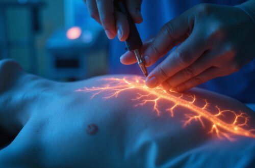
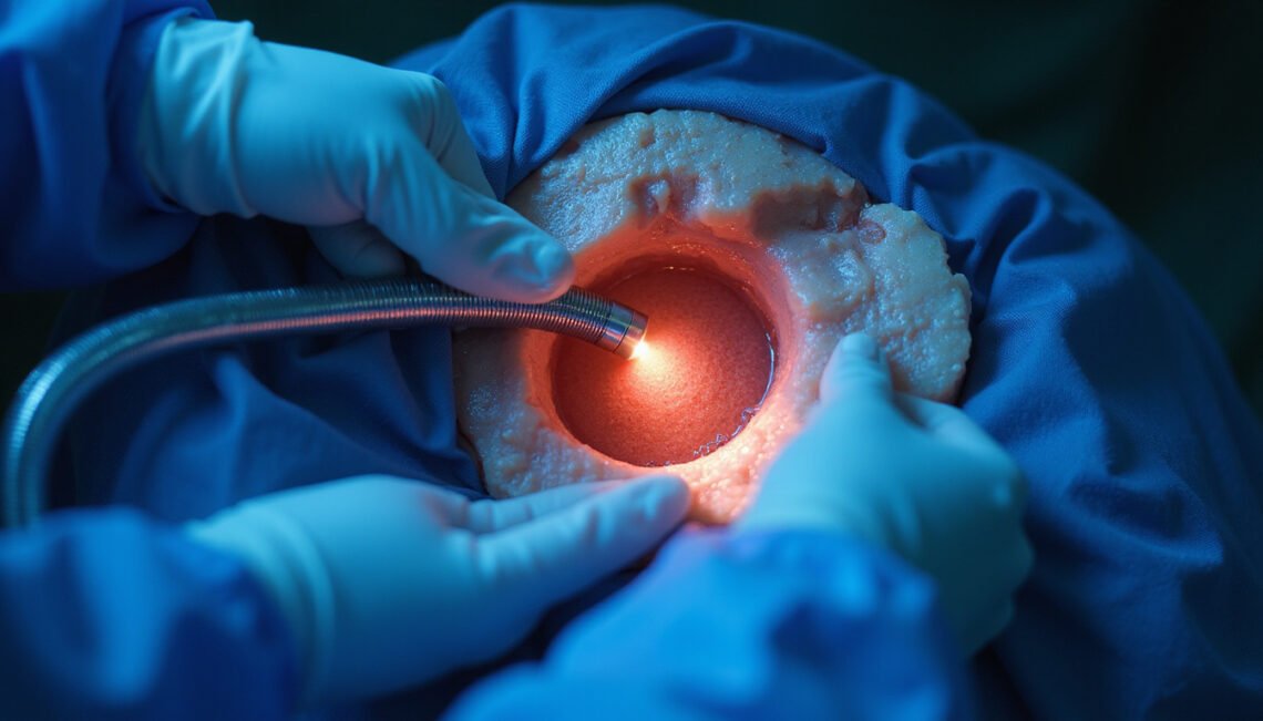
Epiduroscopy (sometimes called spinal endoscopy or epidural endoscopy) is a technique that uses a thin, flexible fiber‑optic camera inserted into the epidural space of the spine. The epidural space is the area just outside the protective covering of the spinal cord and nerves.
Through this small camera, a pain specialist can:
- Visually inspect the epidural space
- Identify scar tissue, inflammation, and adhesions
- Deliver medications directly to painful areas
- Mechanically break up scar tissue that may be trapping nerves
Unlike traditional open back surgery, epiduroscopy is performed through a small incision and guided by live imaging and endoscopic video, making it a minimally invasive outpatient procedure in most cases.
Why Chronic Back Pain Can Be So Hard to Treat
Chronic low back pain and sciatica (leg pain from nerve root irritation) affect millions of people worldwide and are a leading cause of disability (source: World Health Organization). The causes can be complex and overlapping:
- Herniated or bulging discs
- Spinal stenosis (narrowing of the spinal canal)
- Post-surgical scar tissue (epidural fibrosis)
- Inflammation around nerve roots
- Degenerative disc disease
- Facet joint arthritis
Standard imaging—MRI, CT, X‑rays—doesn’t always tell the full story. It may show possible pain generators but can’t always reveal subtle adhesions, inflammatory changes, or how much a nerve root is being tethered by scar tissue. That’s where epiduroscopy can play a unique role: it combines diagnosis and treatment in one procedure.
How Epiduroscopy Works
Epiduroscopy is usually performed by an interventional pain specialist or anesthesiologist in a procedure room or operating theater equipped with fluoroscopy (live X‑ray).
Step-by-Step Overview
-
Preparation and anesthesia
- You’ll change into a gown and have an IV placed.
- Most epiduroscopy procedures use local anesthesia at the insertion site plus light sedation so you’re relaxed but can still respond.
-
Accessing the epidural space
- You lie face down or slightly on your side.
- Using fluoroscopic guidance, the physician inserts a small needle near the tailbone (caudal approach) or occasionally in the lumbar region.
-
Catheter and endoscope insertion
- A flexible catheter is threaded through the needle into the epidural space.
- The tiny fiber‑optic endoscope travels through this catheter, transmitting real‑time images to a monitor.
-
Diagnostic inspection
- The physician slowly advances the scope, visually inspecting the epidural space and nerve roots.
- They may identify scar tissue, inflamed tissue, adhesions, or areas where dye doesn’t spread normally, indicating blockage.
-
Therapeutic interventions
Through the same catheter, the physician can:- Mechanically break up adhesions or scar tissue (adhesiolysis)
- Flush the epidural space with saline to clear inflammatory mediators
- Inject targeted medications such as local anesthetics, steroids, or hyaluronidase
- Occasionally place additional therapeutic catheters if needed
-
Completion and recovery
- The endoscope and catheter are removed, and a small bandage is applied.
- You’ll typically stay in recovery for an hour or two for monitoring, then go home the same day with instructions and a companion to drive you.
Conditions That Epiduroscopy Can Help Address
Epiduroscopy isn’t a cure‑all, but it can be particularly helpful when pain is linked to specific types of pathology within the epidural space.
Common indications include:
-
Post‑surgical back pain (failed back surgery syndrome)
When scar tissue forms around spinal nerves after surgery, it can tether or compress them, causing persistent or recurrent pain. -
Epidural fibrosis
Dense scar tissue in the epidural space that may interfere with nerve gliding and blood flow. -
Chronic radicular pain (sciatica)
Radiating pain, numbness, or tingling down the leg caused by irritated or inflamed nerve roots. -
Adhesions after epidural procedures
Scarring from prior epidural injections or trauma. -
Diagnostic uncertainty
When imaging and clinical findings don’t fully explain the pain, epiduroscopy can offer direct visualization to clarify the diagnosis.
Not everyone with back pain is a candidate for epiduroscopy. It’s generally considered after conservative approaches—physical therapy, medications, standard epidural injections—have been tried without sufficient relief.
Benefits of Epiduroscopy Compared to Other Treatments
For the right patient, epiduroscopy offers several potential advantages:
-
Direct visualization of pathology
Instead of guessing based on MRI alone, the physician can see scar tissue, inflammation, and adhesions in real time. -
Targeted therapy
Medication is placed exactly where it’s needed, which may improve effectiveness compared to blind or fluoroscopy‑only injections. -
Adhesiolysis (breaking up scar tissue)
The mechanical disruption of epidural adhesions is something traditional epidural injections can’t accomplish. -
Minimally invasive
No large incisions, no muscle stripping, and typically no hardware. Recovery is usually much faster than with open spine surgery. -
Diagnostic and therapeutic in one
The procedure can clarify what’s causing your pain and treat it in the same session. -
Surgical delay or avoidance
For some patients, successful epiduroscopy can postpone or eliminate the need for a more invasive spinal operation.
Risks and Possible Complications
Although epiduroscopy is minimally invasive, it is still a spinal procedure and carries some risks. Discuss these in detail with your specialist.
Potential complications include:
- Temporary increase in pain or soreness at the insertion site
- Headache (including post‑dural puncture headache if the dura is inadvertently pierced)
- Bleeding or hematoma
- Infection (e.g., epidural abscess, meningitis—rare but serious)
- Dural tear with cerebrospinal fluid leakage
- Nerve irritation, numbness, or weakness (usually transient)
- Allergic reaction to contrast dye or medications
In experienced hands, serious complications are uncommon, but proper patient selection and aseptic technique are critical.
Who Is a Good Candidate for Epiduroscopy?
Your pain specialist may consider epiduroscopy if:
- You have chronic low back and/or leg pain lasting more than 3–6 months
- Conservative treatments (physiotherapy, medications, standard injections) haven’t provided lasting relief
- Imaging suggests epidural fibrosis, post‑surgical scarring, or nerve root involvement
- You’ve had previous spine surgery and continue to have significant pain
- Surgery has been proposed, but you and your doctor want to explore less invasive options first
You may not be a candidate if you have:
- Uncontrolled bleeding disorders or are on certain anticoagulants
- Active infection, especially near the proposed insertion site
- Severe spinal instability that truly requires surgical stabilization
- Certain severe neurological deficits that demand urgent surgery
A thorough evaluation, including physical exam, imaging review, and sometimes diagnostic nerve blocks, helps determine whether epiduroscopy makes sense in your case.

What to Expect Before, During, and After the Procedure
Before Epiduroscopy
- Medical review: Your doctor will review your medications, allergies, and medical history.
- Medication adjustments: You may need to stop certain blood thinners or diabetes medications beforehand.
- Fasting: Often you’ll be asked not to eat or drink for several hours before the procedure.
- Consent: The risks, benefits, and alternatives will be explained, and you’ll sign a consent form.
During the Procedure
- Time in the procedure room is usually 30–90 minutes.
- You’ll feel local anesthetic at the entry site, then pressure as instruments are advanced.
- With light sedation, you may feel some movement or odd sensations, but you shouldn’t feel sharp pain. Let your doctor know if you do.
After Epiduroscopy
- You’ll be monitored until sedation wears off.
- Mild soreness at the insertion site and temporary changes in pain are common.
- Your doctor may ask you to track pain scores over the following days and weeks.
- Most people resume light activities within a day or two, but heavy lifting and strenuous exercise are usually restricted for a short period.
Pain relief may be:
- Immediate: from the anesthetic and decompression
- Delayed: as steroids or anti‑inflammatory effects build over days to weeks
- Variable in duration: some patients get months to years of relief; others may need repeat interventions or additional therapies
How Effective Is Epiduroscopy?
Results vary, but many studies and clinical experiences suggest that epiduroscopy can significantly reduce pain and improve function in selected patients, especially those with epidural fibrosis or failed back surgery syndrome.
Key points about effectiveness:
- Best outcomes are often reported when there is clear evidence of adhesions or fibrosis corresponding to the pain distribution.
- Combining epiduroscopy with a comprehensive pain management program (physical therapy, exercise, ergonomics, cognitive‑behavioral strategies) usually leads to better long‑term results than the procedure alone.
- Some people require repeat procedures if symptoms recur, while others achieve durable relief after a single intervention.
Your own response will depend on your specific spinal anatomy, pain drivers, overall health, and adherence to rehabilitation recommendations.
Questions to Ask Your Doctor About Epiduroscopy
If you’re considering this procedure, use these questions as a starting point:
- Why do you believe epiduroscopy is appropriate in my case?
- What specific findings are you looking for in my epidural space?
- How many of these procedures have you performed, and what are your typical outcomes?
- What risks apply particularly to me given my health conditions and medications?
- How will this fit into my broader treatment plan—before and after the procedure?
Bringing a family member or friend to your consultation can help you remember details and feel supported as you make decisions.
Quick Summary: Pros and Cons of Epiduroscopy
Potential advantages
- Minimally invasive
- Direct visualization of the epidural space
- Targeted medication delivery
- Ability to break up scar tissue and adhesions
- Often outpatient with relatively quick recovery
- May delay or reduce need for more extensive surgery
Potential disadvantages
- Not suitable for all causes of back pain
- Benefits can be temporary or incomplete
- Requires specialized equipment and expertise
- Carries small but real risks of infection, bleeding, nerve irritation, or dural puncture
- Often part of a larger pain management plan, not a stand‑alone cure
FAQ: Epiduroscopy and Chronic Back Pain
1. How is epidural endoscopy different from a regular epidural injection?
Traditional epidural injections rely on X‑ray guidance but don’t allow direct visualization inside the epidural space. With epiduroscopy, a camera lets the doctor see scar tissue and adhesions, mechanically disrupt them if needed, and place medications precisely where the nerves are irritated.
2. Is spinal epiduroscopy painful or risky?
Most patients experience only mild discomfort during spinal epiduroscopy thanks to local anesthetic and sedation. As with any spinal procedure, there are risks—such as infection, bleeding, or nerve irritation—but serious complications are rare in experienced hands. Your doctor will weigh these risks against the potential benefits for your specific condition.
3. Can endoscopic epidural procedures replace back surgery?
Endoscopic epidural procedures like epiduroscopy can sometimes delay or reduce the need for open back surgery, especially in cases of epidural fibrosis or failed back surgery syndrome. However, they cannot correct major structural problems like severe spinal instability or large disc herniations compressing the spinal cord, which may still require surgical intervention.
Take the Next Step Toward Relief
You don’t have to accept chronic back pain as your “new normal,” nor rush into major surgery without exploring minimally invasive options. Epiduroscopy offers a unique blend of diagnosis and treatment that may uncover hidden sources of pain and address them directly from within the spine.
If your pain has persisted despite conservative care—or if you’ve had spine surgery and still struggle with back or leg pain—consider discussing epiduroscopy with a board‑certified interventional pain specialist. An individualized consultation, review of your imaging, and a clear explanation of risks and benefits can help you decide whether this minimally invasive solution is the right step toward reclaiming your mobility and quality of life.