Nerve Mapping Unlocks Hidden Pain Pathways for Faster Recovery
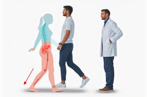
Nerve mapping is transforming how clinicians understand, diagnose, and treat pain. Instead of guessing where pain originates based on symptoms alone, nerve mapping lets specialists “see” the wiring of your nervous system and trace pain to its true source. By identifying hidden pain pathways, this technique can shorten time to diagnosis, guide more precise treatment, and support faster, more complete recovery.
In this guide, you’ll learn what nerve mapping is, how it works, when it’s used, and how it may help you or someone you care about get out of pain sooner.
What Is Nerve Mapping?
Nerve mapping is a set of diagnostic techniques used to identify, trace, and evaluate the function of specific nerves and their branches in the body. It helps clinicians:
- Pinpoint which nerves are irritated, compressed, or damaged
- Distinguish nerve-related pain from muscle, joint, or vascular pain
- Plan targeted interventions such as injections, nerve ablations, or surgery
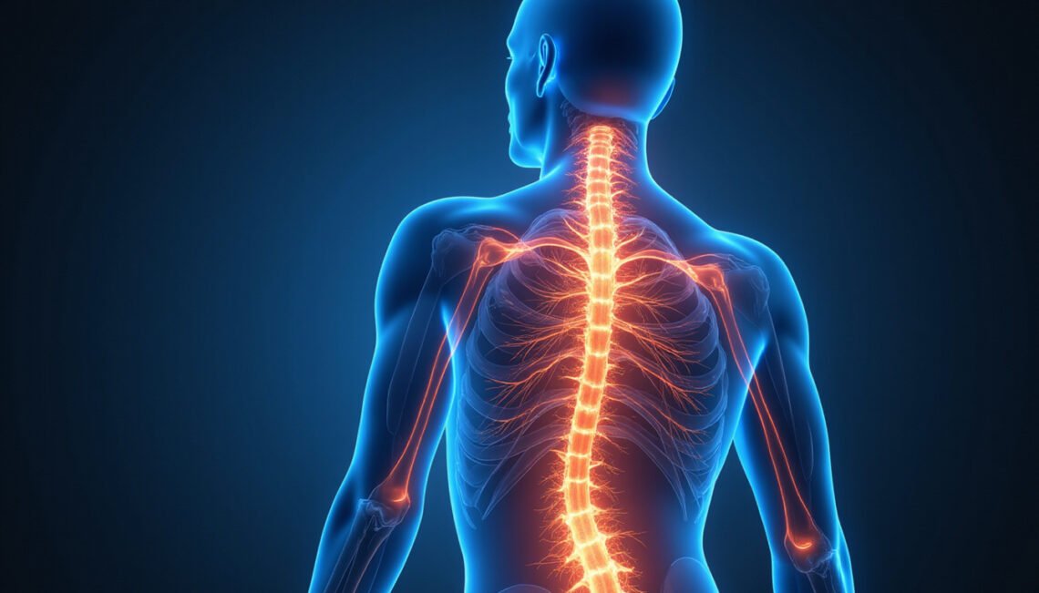
Think of your nervous system as a complex electrical grid. When lights flicker in one room, you can guess which circuit is involved—but with a circuit diagram and a multimeter, you can know. Nerve mapping is that “diagram and multimeter” for the body: it converts vague pain complaints into a precise map for treatment.
Why Traditional Pain Assessment Often Falls Short
Many people with chronic pain spend months or years cycling through medications, physical therapy, and procedures with limited relief. A big reason is that pain can be:
- Referred: felt in one area even though the problem is elsewhere
- Diffuse: spread over a broad region, making it hard to localize
- Multifactorial: involving muscles, joints, discs, nerves, and even the brain
Standard tools—clinical exams, X-rays, or even MRIs—don’t always show which nerve is responsible. An MRI might reveal several disc bulges or arthritic changes, but those findings are common even in people without pain. Treating what “looks bad” on imaging instead of what’s actually generating pain can lead to:
- Unnecessary or ineffective surgeries
- Generalized injections that miss the true source
- Long-term reliance on pain medication without addressing root causes
Nerve mapping aims to cut through this ambiguity by showing which nerves are actively misfiring or irritated right now, not just what structures look abnormal.
How Nerve Mapping Works: The Main Techniques
There isn’t a single, universal method labeled “nerve mapping.” Instead, clinicians use several complementary tools to map nerve function and pain pathways.
1. Electrodiagnostic Testing (EMG and NCS)
Electrodiagnostic testing, which includes electromyography (EMG) and nerve conduction studies (NCS), is one of the most established forms of nerve mapping.
- Nerve Conduction Studies (NCS): Small electrical pulses are applied to a nerve, and sensors measure how quickly and effectively the signal travels.
- Electromyography (EMG): A very thin needle electrode is inserted into muscles to evaluate how well the nerve is activating those muscles.
These tests can help map:
- Which nerve roots or peripheral nerves are affected
- Whether the problem is in the nerve, muscle, or junction between them
- How severe the nerve injury is and whether it’s improving
The American Association of Neuromuscular & Electrodiagnostic Medicine notes that these tests are key for diagnosing conditions like radiculopathy, peripheral neuropathy, and entrapment neuropathies (source: AANEM).
2. Image-Guided Nerve Mapping
Modern imaging has made it possible to visualize nerves and guide targeted diagnostic procedures:
- Ultrasound-guided nerve mapping: High-resolution ultrasound shows superficial nerves in real time. A clinician can track a nerve’s course and perform diagnostic nerve blocks with precision.
- Fluoroscopy- or CT-guided injections: For deeper structures (e.g., spinal nerve roots, facet joints), imaging guides needle placement to the suspected pain generator.
By selectively numbing individual nerves or nerve branches, clinicians can infer:
- “If numbing this nerve relieves your pain, this nerve is involved.”
- “If numbing it doesn’t help, we look elsewhere.”
This process creates a functional map of which nerves actually mediate the pain.
3. Intraoperative Nerve Mapping
Surgeons often use nerve mapping during operations to avoid injury and improve surgical accuracy, especially in:
- Spine surgery
- Neurosurgery
- Orthopedic procedures near major nerves
- Head and neck surgeries
Intraoperative nerve monitoring may involve:
- Stimulating a nerve and observing muscle responses
- Recording signals from nerves to ensure they stay intact
For some patients, this can prevent postoperative weakness, numbness, or new pain by helping surgeons preserve critical pathways.
4. Quantitative Sensory Testing and Pain Diagrams
While more indirect, clinicians may use:
- Sensory testing: Checking light touch, temperature, pinprick, and vibration in specific dermatomes (skin areas supplied by individual nerve roots).
- Pain diagrams: Patients shade areas that hurt. The pattern can suggest particular nerves or roots.
Combined with electrodiagnostics and imaging, these tools refine the nerve map and guide treatment.
Conditions That Benefit from Nerve Mapping
Nerve mapping is especially useful when pain is persistent, unexplained, or poorly responsive to generic treatment.

Common scenarios include:
- Radiculopathy (pinched nerves in the spine): Sciatica, cervical radiculopathy, lumbar nerve root compression
- Peripheral entrapment neuropathies: Carpal tunnel syndrome, cubital tunnel syndrome, thoracic outlet syndrome, tarsal tunnel syndrome
- Chronic regional pain: Complex regional pain syndrome (CRPS), chronic post-surgical pain
- Post-injury or post-operative nerve pain: After fractures, joint replacements, or lacerations where nerve damage is suspected
- Diabetic or metabolic neuropathies: Differentiating diffuse nerve damage from focal entrapments that may be treatable
- Unexplained limb weakness or numbness: To determine whether the problem is in the spine, nerve, muscle, or brain
By clarifying which nerves are impaired or hyperactive, nerve mapping helps focus treatment on the real source instead of chasing symptoms.
How Nerve Mapping Unlocks Hidden Pain Pathways
Pain pathways can be surprisingly complex. Nerves can:
- Branch and rejoin
- Overlap in their sensory territories
- Refer pain along their course or to distant regions
For example:
- A compressed lumbar nerve root may cause pain in the buttock, thigh, and leg, but not always in the back.
- Irritation of a small cutaneous nerve can cause burning pain over a seemingly random patch of skin.
- Post-surgical scar tissue can tether nerves, causing pain that seems unrelated to the original surgery site.
Nerve mapping helps:
-
Localize the origin
By methodically testing and blocking candidate nerves, clinicians can identify the primary generator instead of treating every painful area equally. -
Differentiate overlapping problems
Some patients have both joint arthritis and nerve compression. Nerve mapping reveals how much each contributes, which can be critical when deciding whether surgery is appropriate. -
Reveal “silent” contributors
Occasionally, a mildly compressed or tethered nerve that doesn’t show dramatic changes on MRI can be a major pain driver. If a targeted diagnostic block dramatically relieves pain, that nerve becomes a focus for treatment—even if imaging looks relatively benign.
The Nerve Mapping Process: What to Expect as a Patient
Experiences vary by clinic and technique, but a typical nerve mapping journey may include:
-
Detailed history and physical exam
- Discussion of symptom onset, pattern, triggers, and prior treatments
- Neurological exam assessing sensation, strength, and reflexes
-
Baseline imaging (if indicated)
- X-ray, MRI, or ultrasound to rule out fractures, tumors, or major structural issues
-
Electrodiagnostic testing
- EMG/NCS performed in a clinic setting
- Mild electrical stimulation on the skin; thin needle electrodes in selected muscles
- Takes 30–90 minutes depending on complexity
-
Targeted diagnostic nerve blocks (if needed)
- Performed under ultrasound, fluoroscopy, or CT guidance
- Local anesthetic injected around specific nerve(s)
- Immediate or short-term pain changes are carefully tracked
-
Interpretation and mapping
- All findings (exam, imaging, electrodiagnostics, block responses) integrated into a functional nerve map
- This map guides a personalized treatment plan
-
Follow-up and treatment adjustments
- Reassessment after interventions to see which pathways are resolving
- Map can be updated as symptoms evolve or improve
How Nerve Mapping Supports Faster Recovery
Nerve mapping doesn’t cure pain by itself. Its value lies in shaping what you and your care team do next. Faster recovery comes from better decisions, made earlier:
-
More precise interventions
- Targeted nerve blocks, radiofrequency ablation, or neuromodulation at specific pain generators
- Surgery more narrowly focused on the true culprit nerve or level
-
Avoiding unnecessary procedures
- If mapping shows a nerve is healthy, you may avoid surgeries or injections that were unlikely to help.
-
Optimized rehabilitation
- Physical therapy tailored to the specific nerves and muscles involved
- Better protection of vulnerable nerves during exercise and daily activity
-
Clearer prognosis
- Understanding whether damage is reversible or permanent helps set realistic recovery timelines and goals.
By replacing trial-and-error with a strategic, data-driven approach, nerve mapping can shorten the journey from “What’s wrong?” to “Here’s exactly what we’re going to do about it.”
Potential Risks and Limitations
Like all medical tools, nerve mapping has boundaries:
- Temporary discomfort: EMG needles and nerve stimulations can be mildly uncomfortable. Nerve blocks involve injections.
- Not every pain is nerve-based: Some pain syndromes are more muscular, joint-related, or central (brain and spinal cord level). Nerve mapping may be normal in these cases.
- Interpretation matters: Results are only as good as the clinician’s expertise. Choosing an experienced neurologist, physiatrist, anesthesiologist, or pain specialist is essential.
- No guarantee of cure: Even with a clear nerve map, some chronic pain conditions may be only partially reversible, especially if nerves are severely or long-term damaged.
Despite these limitations, nerve mapping is often a critical piece of an accurate diagnosis for complex pain.
Who Should Consider Nerve Mapping?
You might be a good candidate for nerve mapping if you:
- Have pain, numbness, tingling, or weakness that’s lasted more than a few weeks
- Have had imaging that doesn’t fully explain your symptoms
- Have tried standard treatments (medications, generic PT, basic injections) without lasting relief
- Are considering surgery but want more confidence that the target is correct
- Have pain after surgery or injury that hasn’t been clearly explained
A primary care physician, neurologist, or pain specialist can help decide if nerve mapping is appropriate for your situation.
FAQs About Nerve Mapping and Pain
1. Is nerve mapping painful?
Most people find nerve mapping tolerable. Nerve conduction studies involve brief electrical pulses that may feel like quick taps or zaps. Electromyography uses very fine needles; the sensation is similar to a small injection or pinprick. Image-guided nerve blocks may cause brief discomfort at the injection site, followed by numbness. Your clinician can adjust techniques to minimize discomfort.
2. How long does nerve mapping for back and leg pain take?
Back and leg pain evaluations often combine EMG/NCS with selective nerve root or peripheral nerve blocks. The electrodiagnostic portion typically takes 30–60 minutes. Each guided injection session may add another 30–60 minutes. The full process may unfold over multiple visits to allow accurate interpretation of your responses.
3. What’s the difference between nerve mapping and nerve conduction studies?
Nerve conduction studies are one component of nerve mapping. Nerve mapping is a broader concept that includes:
- Nerve conduction studies
- Electromyography
- Imaging and image-guided injections
- Sensory exams and pain pattern analysis
- Intraoperative monitoring (when applicable)
Together, these create a comprehensive “map” of nerve function and pain pathways, whereas nerve conduction studies alone focus on how electrical signals travel along specific nerves.
Take the Next Step Toward Accurate Diagnosis and Faster Relief
Living with persistent pain can feel like being lost without a map—every new treatment is another guess, and progress is slow. Nerve mapping gives you and your care team a clear, evidence-based view of your pain pathways, so decisions are targeted instead of trial-and-error.
If your pain has outlasted simple treatments, or if you’re facing a major decision like surgery, ask your healthcare provider whether nerve mapping techniques—such as EMG, nerve conduction studies, or image-guided diagnostic blocks—are right for you. Seeking an evaluation with a specialist who routinely uses nerve mapping can be the turning point that unlocks hidden pain sources and puts you on a faster, more confident path to recovery.